When you ask what do hair follicles look like, you’re really seeking a clear picture of the tiny but complex structures that generate every strand of hair on your scalp. Understanding the appearance of these follicles helps you recognize a healthy scalp, identify potential problems, and appreciate the precision involved in modern hair‑restoration procedures.
Every year, millions of people worldwide consider hair transplantation as a solution for thinning hair. Yet, many are unfamiliar with the microscopic anatomy that surgeons work with during a follicular unit extraction (FUE) or direct‑hair‑implantation (DHI) session. By visualizing the follicle’s components—such as the hair bulb, hair papilla, and hair shaft—you gain confidence in the process and can ask informed questions.
In this article we will explore the detailed anatomy of a follicle, how its appearance changes throughout the hair growth cycle, what common conditions can alter its look, and what surgeons actually see when they extract a graft for transplant. Let’s dive into the science that underpins a full, natural‑looking head of hair.
Understanding the Structure of a Hair Follicle
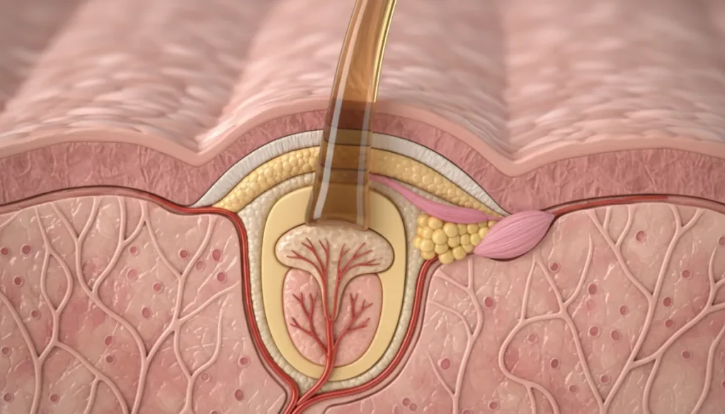
The hair follicle is a dynamic mini‑organ embedded in the skin that produces the visible hair strand. From the surface of the scalp down to the deeper layers of the dermis, each follicle consists of several distinct parts, each with a specific function.
Key Components
- Hair Bulb: The rounded base at the bottom of the follicle where cells actively divide.
- Hair Papilla: A cluster of specialized mesenchymal cells that supply nutrients and signals to the bulb.
- Hair Shaft: The part that extends outward, composed of keratinized cells that become the visible hair.
- Hair Root: The portion of the shaft located below the skin surface, anchored within the follicle.
- Outer and Inner Root Sheaths: Protective layers that guide and shape the growing shaft.
Below is a simple comparison that highlights the visual differences between these structures when observed under a microscope or during a transplant extraction.
| Component | Location | Typical Appearance |
|---|---|---|
| Hair Bulb | Base of follicle | Rounded, pigmented, slightly enlarged |
| Hair Papilla | Within bulb | Light‑colored, vascular, appears as a tiny “button” |
| Hair Shaft | Extends outward | Long, cylindrical, varies in thickness |
| Root Sheath | Surrounds shaft | Layered, translucent, protective |
When a surgeon extracts a graft, the visible portion usually includes the hair shaft up to the bulb, with the papilla intact. This is the exact visual that patients often wonder about when they ask, “what do hair follicles look like after extraction?”
How Hair Follicles Appear at Different Growth Stages
The look of a follicle changes dramatically as it moves through the three primary phases of the hair growth cycle: anagen (growth), catagen (regression), and telogen (rest). Recognizing these visual cues helps clinicians assess scalp health and plan effective transplant strategies.
Anagen – The Active Growth Phase
During anagen, the follicle is at its largest. The hair bulb swells, the papilla is richly vascularized, and the hair shaft elongates rapidly. Under magnification, an anagen follicle appears robust, with a dark‑pigmented bulb and a clear, thickening shaft.
Catagen – Transitional Regression
In catagen, the follicle shrinks. The bulb contracts, the papilla loses some vascularity, and the shaft stops elongating. Visually, the bulb becomes smaller and lighter, and the surrounding sheath begins to separate, giving the follicle a “pin‑like” appearance.
Telogen – Resting Phase
Telogen follicles are the smallest and often invisible to the naked eye. The bulb has fully retracted, the papilla is dormant, and the shaft is fully formed but detached, awaiting shedding. When observed during a scalp examination, telogen follicles may appear as tiny, pale pits.
The table below summarizes the visual characteristics of each phase:
| Phase | Bulb Size | Papilla Vascularity | Visible Shaft Length |
|---|---|---|---|
| Anagen | Large, pigmented | High | Increasing (up to several centimeters) |
| Catagen | Medium, lighter | Medium | Shortening |
| Telogen | Small, pale | Low | Fully formed, not growing |
Understanding these visual differences is essential for anyone asking what do hair follicles look like at various stages, especially when evaluating donor areas for transplantation.
Common Conditions That Change the Appearance of Follicles
Various scalp disorders can alter the typical look of a follicle, making it appear blocked, inflamed, or even dead. Recognizing these changes is vital for early treatment and for planning a successful hair transplant.
Blocked or Clogged Follicles
Excess sebum, dead skin cells, or bacterial buildup can clog the follicular opening. Visually, a blocked follicle may appear as a tiny, raised bump with a darkened tip. This condition often leads to comedones or acne‑like lesions on the scalp.
Inflamed Follicles (Folliculitis)
When bacteria invade the follicle, inflammation occurs. The affected area becomes red, swollen, and may exude pus. Under a dermatoscope, the follicle looks enlarged with a bright rim of irritation.
Dead or Dormant Follicles
Age, genetics, or severe hormonal imbalance can cause follicles to cease activity. Dead follicles lose pigmentation, shrink, and the papilla appears pale. They may be mistaken for normal telogen follicles, but they no longer produce a viable shaft.
Other Visual Changes
- Ingrown hairs: The shaft curves back into the skin, creating a raised, sometimes painful nodule.
- Scarring alopecia: Fibrotic tissue replaces normal follicular structures, resulting in a smooth, scarred patch.
- Hair shaft disorders (e.g., pili torti): The shaft twists, giving the follicle a corkscrew appearance.
By learning what do hair follicles look like under these conditions, patients can better discuss symptoms with dermatologists or transplant surgeons and pursue appropriate interventions.
What to Expect When a Follicle Is Extracted for Transplant
During an FUE or DHI procedure, the surgeon isolates individual follicular units that typically contain 1‑4 hairs. The visual of each extracted graft is a key concern for many patients asking, “what do hair follicles look like after they are pulled out?”
Step‑by‑Step Visual Guide
- Identification: Using magnification, the surgeon locates a healthy anagen follicle with a robust bulb.
- Incision: A micro‑punch (0.8‑1.0 mm) creates a tiny circular cut around the follicle.
- Extraction: The graft is gently lifted, preserving the hair papilla and surrounding sheath.
- Inspection: Under a stereomicroscope, the graft is examined; a healthy graft appears as a slender shaft ending in a dark‑pigmented bulb with a visible papilla.
- Storage: The graft is placed in a chilled solution to maintain viability until implantation.
Visually, a well‑extracted follicle resembles a tiny, elongated “pearl” with a dark tip (the bulb) and a thin, translucent shaft. Any damage to the papilla or excessive stripping of the sheath can compromise graft survival, which is why precise visual assessment is critical.
Understanding the exact look of a harvested follicle helps patients set realistic expectations for graft survival rates and post‑procedure results.
Why Choose Gold City Hair
At Gold City Hair, our team combines years of expertise with the latest FUE and DHI technologies to ensure each graft is harvested and implanted with meticulous care. Since 2017, we have helped thousands of clients restore confidence through natural‑looking results, transparent communication, and personalized treatment plans. Our clinic in Turkey is dedicated to delivering safe, effective hair restoration while prioritizing your comfort and satisfaction.
Ready to see the difference a healthy follicle can make? Contact Gold City Hair today for a personalized consultation and take the first step toward a fuller, more confident you.
FAQ
What do hair follicles look like?
Hair follicles are tiny tube‑like structures embedded in the scalp that consist of a bulb, papilla, shaft, and surrounding sheaths.
How does a hair follicle appear during the anagen (growth) phase?
During anagen the follicle is at its largest, with a dark‑pigmented bulb, a highly vascular papilla, and a thickening shaft.
What visual changes occur in a hair follicle during the catagen (regression) phase?
In catagen the follicle shrinks, the bulb becomes smaller and lighter, and the papilla loses some vascularity.
How can a blocked or inflamed hair follicle be recognized?
Blocked follicles appear as tiny raised bumps with dark tips, while inflamed follicles show redness, swelling, and sometimes pus.
What does a hair graft look like after it is extracted for a transplant?
A harvested graft appears as a slender shaft ending in a dark‑pigmented bulb with an intact papilla, resembling a tiny pearl.
Why is the appearance of the hair papilla important in hair transplants?
The papilla supplies nutrients to the bulb; a healthy papilla ensures graft viability and successful hair growth after transplantation.